

Peri-Implantitis: What It Is, Why It’s So Common, and How We Keep It From Starting
By Dr. Niels Oestervemb, DDS — Double Board-Certified: Implant Dentistry (ABOI/ID) & General Dentistry (ABGD) · Zero Bone Loss Concepts Ambassador
Peri-implantitis is an infection of the gum and bone around a dental implant that destroys the bone holding it. It is the leading reason implants fail years after a surgery that was called a success. In large published reviews, roughly one in five implant patients develops it within five to ten years, and close to half show the milder, reversible early stage at some point. It is nearly painless until it is advanced — and most of it is preventable through decisions made before the implant ever goes in.
Two stages, one direction
- Peri-implant mucositis — inflammation of the gum around the implant with no bone loss yet. Bleeding when probed or brushed, some redness or puffiness. Reversible with professional cleaning and better home care.
- Peri-implantitis — the same inflammation, now with progressive loss of the bone around the implant. The gum pocket deepens, pus may appear, threads become exposed. Bone that is lost does not grow back on its own. Treatable, but rarely fully reversible.
The difference between the two is time. Mucositis that nobody catches becomes peri-implantitis; peri-implantitis that nobody catches becomes a loose implant.
Symptoms — and why most people have none
Bleeding around the implant when you brush or when it is probed. Redness, swelling, or a gum that looks puffier than its neighbors. A bad taste or odor from the site. The gum pulling back so a grey collar or metal threads start to show. Pus if you press on the gum. Later: tenderness when chewing, and — at the end — an implant that moves.
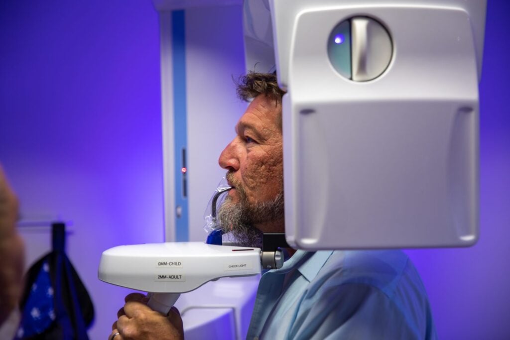
Notice what is missing from the top of that list: pain. Implants have no ligament and no nerve supply of their own, so the warning system natural teeth give you is absent. Bleeding is the smoke alarm. By the time an implant hurts, the bone loss is usually years old. That is why a yearly bone-level X-ray is not optional for anyone with implants, and why we offer a free bone-level check for implants placed anywhere.
What causes it
Bacteria cause the infection. But bacteria are in every mouth; what decides whether they win is the environment the implant was given. The risk factors fall into two groups.
Decided before and during treatment — by the dentist:
- Thin gum tissue. Implants placed into tissue under 3 mm thick lose bone in the first year to make room for a biological seal; thicker tissue does not. Research from Prof. Tomas Linkevičius’ group measured the difference at up to 1.45 mm of loss in thin tissue against about 0.2 mm in thick. Bone lost that early is the pocket that peri-implantitis later moves into.
- Cement left under the gum. Cement-retained crowns leave excess cement in the gum pocket more often than anyone intends, and leftover cement is one of the best-documented triggers of peri-implantitis there is. Screw-retained teeth have no cement at all.
- Placement depth and position. An implant placed too deep, too shallow, or angled where it cannot be cleaned is an implant that will collect plaque where no brush reaches.
- The seam. A loose implant-to-abutment connection pumps bacteria at the exact level of the bone. A tight conical connection and a polished collar give bacteria far less to colonize.
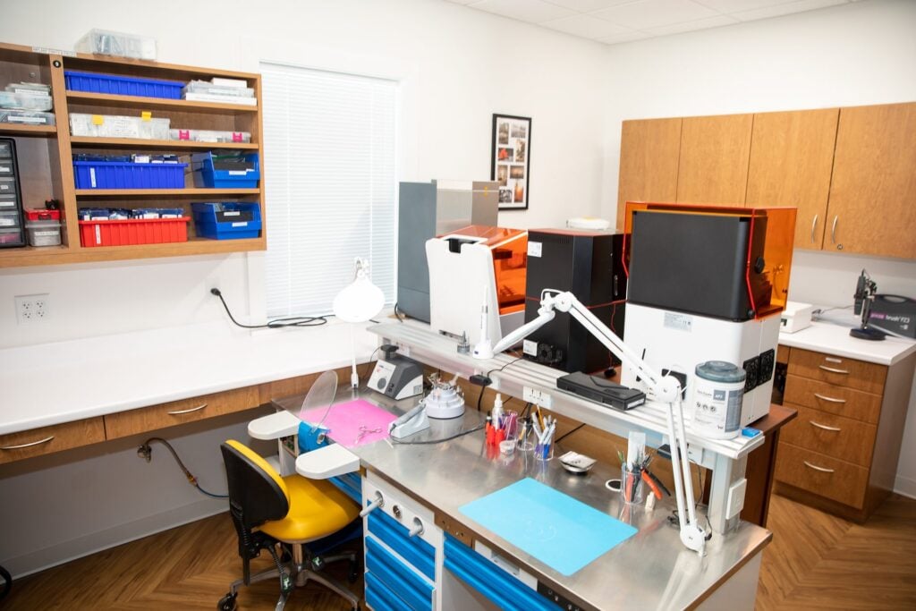
- The shape of the tooth. An over-bulked crown or bridge that flares out at the gumline traps plaque and blocks cleaning. The contour where the tooth emerges from the gum — the emergence profile — is designed, or not, in the lab.
Decided after treatment — by the patient:
- A history of gum disease. The bacteria that took natural teeth are still there. Implants in a mouth with untreated periodontitis are the highest-risk implants we see.
- Smoking — roughly doubles the risk of implant failure in most studies and blunts every treatment.
- Uncontrolled diabetes, which feeds infection and slows the body’s response.
- Home care and maintenance. Implants do not decay, so people stop worrying about them. The bacteria do not stop.
- Grinding. Overload does not cause the infection, but it accelerates bone loss once infection starts. A night guard is cheap insurance.
Why the profession accepts so much of this
The success criteria dentistry has used since 1986 accept up to about 1.5 mm of bone loss around an implant in the first year of function, then 0.2 mm every year after. An implant can meet that definition of “success” while quietly building the pocket that peri-implantitis will move into. We think that standard is out of date, and so does the research — Dr. Oestervemb trained with Prof. Linkevičius, the researcher who showed that early bone loss is preventable, and follows his protocol on every implant placed here.
Treatment — what works, and what to be honest about
Mucositis (no bone loss yet): professional cleaning of the implant surface, a corrected home-care routine, and a recheck in a few weeks. This stage is reversible, and catching it is the single most valuable thing a maintenance visit does.
Early peri-implantitis (shallow pockets, little bone lost): thorough non-surgical decontamination of the implant surface, often with a short course of antibiotics, then tight follow-up. Many of these cases stabilize.
Moderate peri-implantitis: non-surgical cleaning cannot reach the bottom of a deep, infected pocket, so the gum is opened to clean and decontaminate the implant directly. Where the bone defect is contained — a crater around the implant — regenerative grafting can rebuild part of what was lost. Where it is not, exposed threads are smoothed and polished (implantoplasty) so plaque has nowhere to hold, and the gum is reshaped so you can clean it. Often the crown or bridge is redesigned at the same time, because a restoration that caused the problem will cause it again.
Advanced peri-implantitis: when most of the supporting bone is gone or the implant moves, the honest treatment is removal, grafting of the site, healing, and in most cases a new implant placed with the variables controlled this time. How we rescue and replace failing implants is covered here.
The honest part: no treatment for established peri-implantitis is as reliable as never developing it. Published results for surgical treatment are respectable but imperfect, recurrence is common when risk factors are not changed, and cost rises steeply at each stage — from a deep cleaning, to several thousand dollars for surgical regeneration, to more than the original implant cost for removal and replacement. Rarely in dentistry is procrastination priced so steeply.
How we keep it from starting
Every implant placed here follows a published six-step standard: gum thickness measured before surgery and thickened if under 3 mm; placement at the depth the implant’s design calls for, planned in 3D and fully guided; implants with a tight conical connection and a polished collar; screw-retained zirconia teeth polished below the gumline with no cement, ever; the emergence profile designed in our own lab; and a bone-level X-ray at delivery, rechecked at every follow-up, with the number shown to you. The whole standard, in plain English, is here. Add your half — cleaning the way we teach you, maintenance visits, a night guard if you grind, and not smoking — and the odds swing hard in your favor.
Already have implants? Find out where your bone is.
If your implants were placed anywhere — including here — and you cannot remember the last time someone measured the bone around them, that is the appointment to make. Bring your most recent X-rays or let us take a fresh 3D scan at no charge. Dr. Oestervemb will measure each implant, tell you plainly what he sees, and put it in writing. Stable is a result worth hearing. Starting to move is a result worth hearing early. Book a free implant bone check.
Frequently asked questions
Can peri-implantitis be cured?
The infection can be controlled and the implant kept, especially when it is caught early. Bone that has already been lost is only partly rebuildable, and only in favorable defects, which is why the goal is to catch it at the mucositis stage or prevent it altogether.
Is peri-implantitis reversible?
The early stage — peri-implant mucositis, inflammation without bone loss — is reversible with cleaning and better home care. Once bone has been lost, the disease can be stopped but the lost bone does not return on its own.
Does peri-implantitis hurt?
Usually not until it is advanced. Implants have no ligament and no nerve supply of their own, so bleeding, swelling, a bad taste, or receding gum are the signs to act on — not pain.
How is it different from gum disease?
It is the same kind of bacterial infection, but around an implant instead of a tooth, and it tends to progress faster because an implant lacks the ligament and blood supply that help a natural tooth defend itself. People with a history of gum disease are at the highest risk.
Will I lose the implant?
Not if it is caught early and the causes are corrected. Implants are lost to peri-implantitis mostly when it goes unnoticed for years — the reason a yearly bone-level X-ray matters more than almost anything else you can do for an implant.
How much does peri-implantitis treatment cost?
It depends entirely on the stage: a deep cleaning for the early stage costs a fraction of surgical regeneration, which in turn costs less than removing and replacing an implant. We quote in writing after a scan, and we will tell you when the right answer is to watch rather than treat.
Prevalence figures and treatment descriptions are drawn from published research for education; your diagnosis and options follow an exam.


